 Urology · Educational animation
90 seconds
Histotripsy for a kidney mass
Histotripsy is being studied for kidney masses. Here is how it works.
Ask an LLM to review this video
Copy this prompt into your reviewer. If it cannot open the video, upload the MP4 using one of the links above.
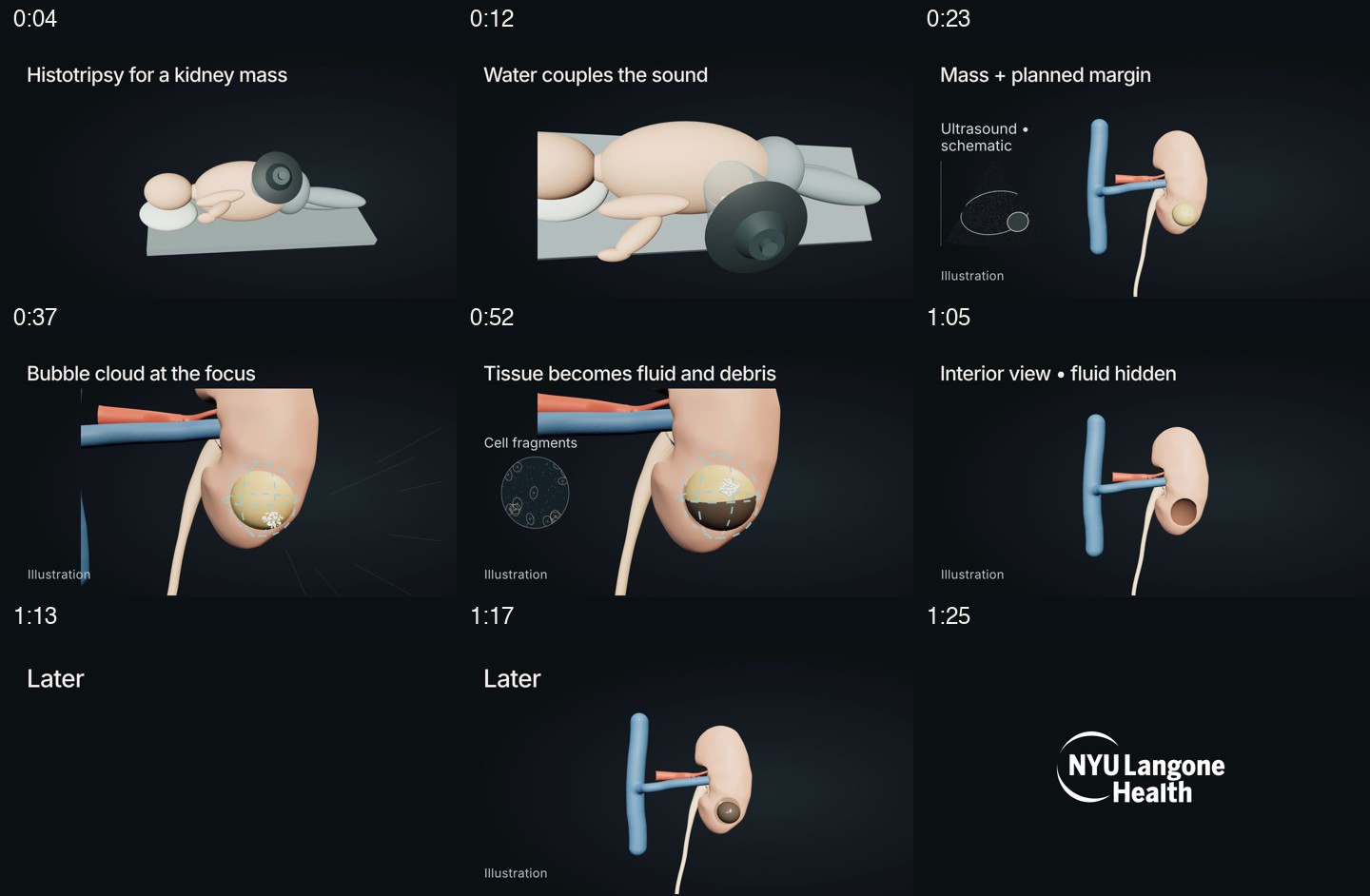
Timestamped video stills · Narration transcript · Clinical sources and limits
Narration transcript
Histotripsy for a kidney mass
90-second narration transcript

0:00–0:08
Histotripsy is being studied for kidney masses. Here is how it works.

0:08–0:18
The patient lies on their side. A water bath against the skin links the treatment head to the body.

0:18–0:29
Ultrasound imaging helps the team locate the mass and plan the treatment area, including a margin around it.

0:29–0:46
Short sound pulses pass through the skin without an incision. They meet at a small focus, where tiny bubbles form, expand, and collapse. This breaks tissue apart mechanically. Heat is not the treatment mechanism.

0:46–0:59
The focus moves through the planned area. Solid tissue breaks down into fluid containing tiny cell fragments, called acellular debris.

0:59–1:12
We hide the fluid in this illustration to show the space inside the treated area. The kidney's outer contour remains visible.

1:12–1:23
Later, the body can clear the broken-down material, and the treated area can shrink. This change takes time.

1:23–1:30
The team uses imaging and clinical assessment to decide whether a kidney mass can be treated.
Clinical sources
The cavity view explicitly hides the fluid as a teaching illustration. “Later” marks a time jump; the film gives no healing timetable or outcome rate.
- Wah et al. CAIN feasibility trial protocol, 2025
Abstract; Histotripsy Device; Index-Procedure Ultrasound Imaging Inclusion/Exclusion; Histotripsy Treatment. Primary renal protocol, not outcome evidence.
- Wah et al. Early Clinical Histotripsy Treatment of Renal Cell Carcinoma, 2025
Treatment-position and planning paragraphs; later imaging paragraph; procedural considerations. One renal case, not a rate or general prognosis.
- NYU Langone: Histotripsy Makes Its Clinical Debut in NYC
Opening mechanism paragraph. Describes cavitation, acellular debris and reabsorption. Clinical service described is liver; do not transfer its outcomes or recovery claims to kidney.
- Hall et al. Histotripsy of rabbit renal tissue in vivo: temporal histologic trends, 2007
Abstract results/conclusions. Animal renal tissue; later resorption, fibrosis and a contour defect were reported. No human timing or unchanged-contour guarantee.
- NYU renal histotripsy research listing
Brief description; trial identifier NCT05820087. Supports renal research context only.
- NCI renal histotripsy trial listing
Description; status closed to accrual. Guard against an unsupported recruitment invitation. Trial and device names stay in sources, outside the film.